Biomechanics
Biomechanical engineers apply all aspects of civil engineering including engineering principles and techniques into the medical field. Our research group provides new and innovative solutions utilizing sophisticated tools to help solve various biomechanics research problems. Our biomechanics lab is leading in a variety of multidisciplinary areas in human biomechanics related to musculoskeletal system and human topography.
Our work in the biomechanics of musculoskeletal system includes but not limited to Finite Element (FE) modelling of the human spine in both lumbar and cervical region, FE modelling of diarthrodial joints (hip, knee and ankle joints) with application in designing artificial joint implants as well as modeling and designing human assistive equipment. Our lab is also one of the leaders in implementing body/part topography in clinically relevant issues such as implementing modern three dimensional mapping techniques to study the variation of the geometry of diarthrodial joints (ankle joint) with implant design application as well as implementing three dimensional surface scanning techniques as non-invasive sensitive measures to diagnose and monitor the scoliotic condition. Our research projects are conducted both computationally and experimentally associated together with our latest software and hardware facilities.
Computational Abilities
The computational parts of our projects are mainly performed by means of different softwares including Mimics, Geomagic, Hypermesh, Abaqus, etc., as well as in-house programs implemented in Matalab or Mathematica.
Reconstruction of the 3D Geometry
Reconstruction of the body parts are done by segmentation of the CT-Scan/MRI/Ultrasound slides in three anatomical planes. The medical image processing software, Mimics (Materialise, Belgium), is employed to create the 3D geometry.
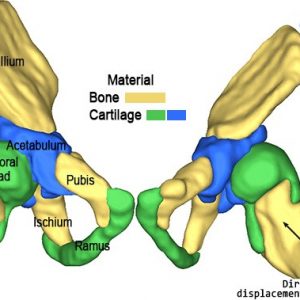
Reconstruction of the infant hip from Ultrasounds; comparison of the normal and dysplastic cases.
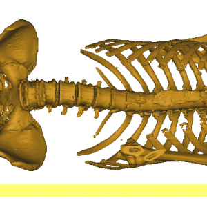
Patient skeleton laying on the left side position, in order to measure pelvic parameters to be used in hip replacement arthroplasty.
Geometrical Analysis
3D geometrical analysis of contours of deviation are implemented in different applications using Geomagic Studio/Control (3D Systems, USA). For example, surface topography asymmetry analysis is employed for classifying, assessing, and monitoring adolescent idiopathic scoliosis. Our surface topography asymmetry maps correspond to the spine deviation observed in radiographs:
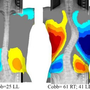
Isolated color patch of two torso overlapped with the corresponding radiographs

The torso deviation color map of patient with adolescent idiopathic scoliosis
Finite Element (FE) Mesh
Stress analysis of the musculoskeletal system usually requires Finite Element (FE) analysis due to its structural complexity. FE mesh (generated via Hypermesh, Altair, USA) of the biomechanical models typically includes 1D, 2D and 3D element connecting and interacting together to simulate different tissues with complex geometries.
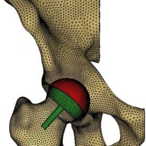
FE mesh of the hip resurfacing arthroplasty.

Micro-scale finite element modeling of ultrasound propagation in aluminum trabecular bone-mimicking phantoms. (a) The finite element model simulating the reference signal (experimental setup without a sample). (b) The finite element model of a computational aluminum foam sample surrounded by the water (acoustic) medium. (c) A view of the generated finite element meshes for the sample and its saturating water medium.
Finite Element (FE) analysis
The FE computations are implemented by Abaqus (Dassault Systems Simulia Corp., USA) to calculate stress/force and strain/deformation of every single element.
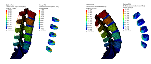
Kinematics of the lumbosacral spine due to flexion/extension movements along with stress distribution in intervertebral discs.

Von Mises and pressure stresses in lizard jaw due to biting forces


